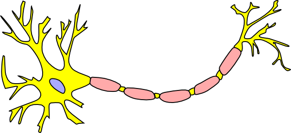
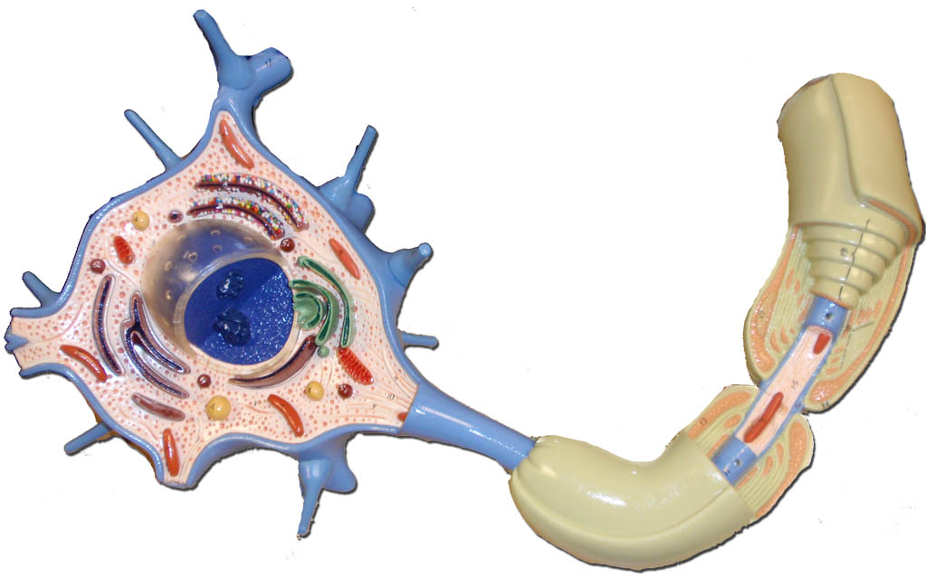
42 picture of a neuron without labels
Types of Neurons: Parts, Structure, and Function - Verywell Health Summary. Neurons are responsible for transmitting signals throughout the body, a process that allows us to move and exist in the world around us. Different types of neurons include sensory, motor, and interneurons, as well as structurally-based neurons, which include unipolar, multipolar, bipolar, and pseudo-unipolar neurons. Nervous System Anatomy Stock Photos And Images - 123RF Affordable and search from millions of royalty free images, photos and vectors. Photos. Vectors. FOOTAGE. AUDIO. SEE PRICING & PLANS. Support. en ... Nervous system. Human anatomy. Brain, motor neuron, glial and.. Vector. Similar Images . Add to Likebox #85341064 - Neurons cells concept ... BLOOD VESSELS_Labels. Similar Images . Add to Likebox ...
Pics Of Labeled Of A Neuron Pictures, Images and Stock Photos Motor neuron, detailed and accurate, labeled. The nervous system The human nervous system vector medical illustration pics of labeled of a neuron stock illustrations. The nervous system. Dendritic cells vector illustration. Anatomical labeled closeup scheme with progenitor, immature, nucleus and membrane extensions.
Picture of a neuron without labels
Neurons: Meaning, Types, Functions, Diagrams - Embibe What is Neuron and its Function? Neurons are the basic and fundamental units of the nervous system which are responsible for transmitting signals to establish communication between the central nervous system and the body. Neurons are also called nerve cells. Neurons use electrical and chemical signals to coordinate all the essential functions of life. ... Labeled Neuron Diagram - Science Trends Neurons are a type of cell and are the fundamental constituents of the nervous system and brain. Neurons take in stimuli and convert them to electrical and chemical signals that are sent to our brain. There are 3 major kinds of neurons in the spinal cord: sensory, motor, and interneurons. Neuron Cell Worksheets - Superstar Worksheets This neuron coloring sheet is perfect for younger students to give them an idea of what a neuron cell looks like and all of their details. Neuron Cell Journaling Page Having a science notebook or journal handy helps reinforce key facts! Plus, use them as coloring pages while writing important key facts on the cell notebooking page.
Picture of a neuron without labels. What Neurons Look Like (as Drawn by Students, Grad Students, and ... According to a new study, your sketch will depend on how much science education you have, but not in the way you'd expect. In the image above, the top row -- those detailed, labeled, neat... › ~kimscott › slides7. Artificial neural networks - MIT without assigning the labels penny, ^nickel, _ ^dime, _ ^quarter _) We will focus on supervised learning. They can also perform ^association _ tasks, for instance reproducing a full image from a small piece. The learning problem If you show a picture to a three-year-old and ask him if there is a tree in it, he is likely to give you the Neuron Diagram Unlabeled neuron, (1). axon, cell body, dendrites, nucleus, terminal. Unlabeled diagram of a motor neuron (try labeling: axon, dendrite, cell body, myelin, nodes of Ranvier, motor end plate).Read the definitions, then label the neuron diagram below. axon - the long extension of a neuron that carries nerve impulses away from the body of the cell. superstarworksheets.com › science-worksheetsPhases of the Moon Worksheets - Superstar Worksheets A shaded chart without labels to use as review of the different phases of the moon as seen from Earth. Phases of the Moon Cut & Paste A cut and paste worksheet for children to arrange the phases of the moon then label them accordingly.
What Is a Neuron? Diagrams, Types, Function, and More Takeaway. Neurons, also known as nerve cells, send and receive signals from your brain. While neurons have a lot in common with other types of cells, they're structurally and functionally unique ... Neuron (Nerve Cell) Types, Structure and Function Neurons, also known as nerve cells, are essentially the cells that make up the brain and the nervous system. Neurons do not touch each other, but where one neuron comes close to another neuron, a synapse is formed between the two. The function of a neuron is to transmit nerve impulses along the length of an individual neuron and across the ... Yvonnes neuropsychology pictures - GLITTRA Below are four pictures that can be used for learning the names of the different parts of the cortex of the brain. They show a lateral view of the gyri of the cortex, a lateral view of the sulci (fissures), a medial view of the gyri and a medial view of the sulci, respectively. Back home! › science › articleWidespread implementations of interactive ... - ScienceDirect May 10, 2022 · For each neuron, we then determined if each time bin had a significant area under the curve (AUC) value by shuffling its firing rates and ROI labels 100 times (p < 0.01, permutation test). Neurons with significant AUC values for at least 5 consecutive bins were included in further analyses and were sorted based on the first bin with a ...
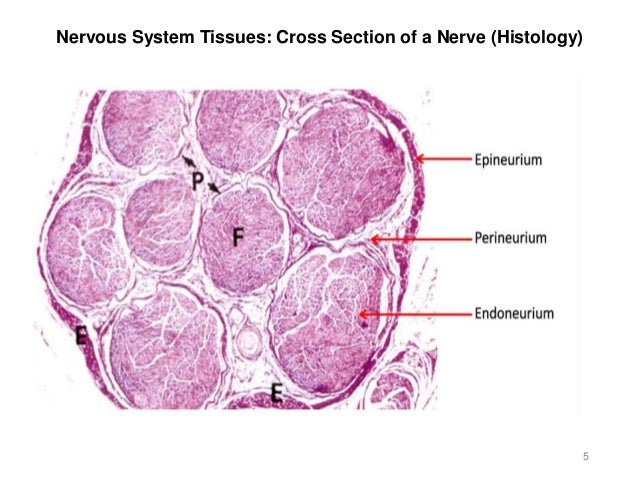
Nervous system Images, Stock Photos & Vectors | Shutterstock Nervous system royalty-free images. 56,172 nervous system stock photos, vectors, and illustrations are available royalty-free. See nervous system stock video clips. Set goals and get predicted insights based on performance. › 43014289 › Neuroscience_by_DaleNeuroscience by Dale Purves et al. (eds.) (z-lib.org) Enter the email address you signed up with and we'll email you a reset link. 5,484 Central Nervous System Stock Photos and Images - 123RF Central Nervous System Stock Photos And Images. 5,484 matches. Page of 55. Sympathetic And Parasympathetic Nervous System. Difference. diagram with connected inner organs and brain and spinal cord. Educational guide of human anatomy. vector illustration for medical and science use. types of neurons: sensory and motor neurons, and interneuron. Neuron Diagram & Types | Ask A Biologist Neurons and Nerves Neurons are unique for many reasons. For one, they have a shape that is not like any other cells. Nerve cells are also some of the longest cells in your body. There are nerve cells as long as a meter. They stretch from your hips all the way down to your toes! This is very uncommon for cells, which are usually very short. Most cells are 20 micrometers in
Index of /~david/courses/perception/lecturenotes/brain/brain-slides
Labeled brain anatomy Images, Stock Photos & Vectors - Shutterstock Images libres de droits de Labeled brain anatomy. 2,779 photos, images vectorielles et illustrations de labeled brain anatomy libres de droits sont disponibles. Afficher les clips vidéos de stock de labeled brain anatomy. Fixez des objectifs pour connaître les résultats attendus.

Neuron Diagram Unlabeled - Wiring Diagram Pictures Find nerve cell diagram Stock Images in HD and millions of other royalty-free stock Related: axon and dendrites, neuron myelin, cell education, neural cells, . Read the definitions, then label the neuron diagram below. axon - the long extension of a neuron that carries nerve impulses away from the body of the cell.

worksheet. Nervous System Worksheet. Grass Fedjp Worksheet Study Site
› Research › monkeylogicMonkeyLogic - Brown University Introduction. MonkeyLogic is a MATLAB toolbox for the design and execution of psychophysical tasks with high temporal precision.It is structured to allow for the flexible construction of sensory, motor, or cognitive tasks that are based upon the interaction of a subject with visual stimuli through the use of eye-position, joystick, button, lever, and / or keyboard input.
7th Science - J. Famigletti: Resources
Label the Parts of a Neuron | Neurons, Cells worksheet ... - Pinterest The images on this identification worksheet have a more basic engineering look than commonly used photographs. This simplicity helps students to understand the six simple machines in their most basic form, and to be able to better recognize them in everyday applications. Free to print (PDF file). S Student Handouts Primary Grades Neuroscience
Activity 7 - Brain & Cranial Nerves
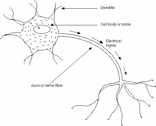
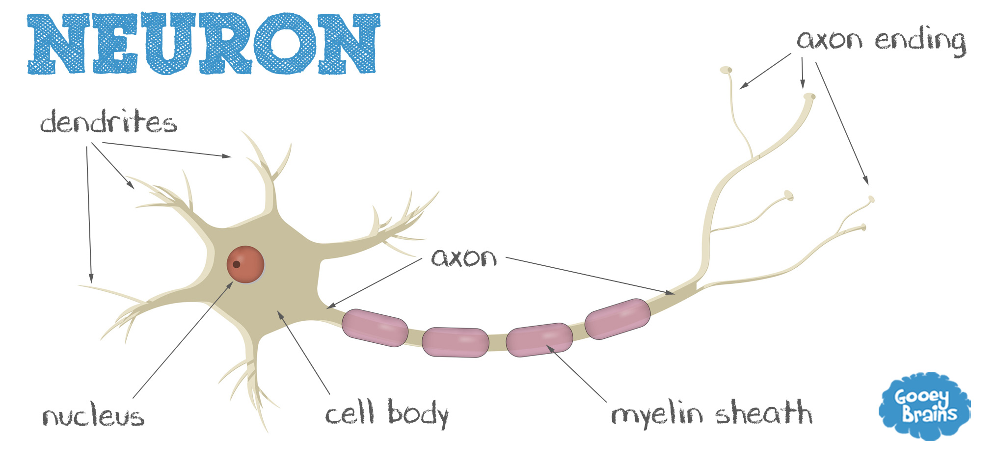
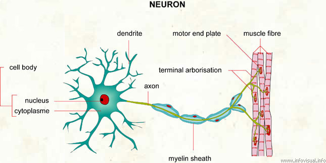
Parts of a Neuron and How Signals are Transmitted - Verywell Mind Axon. The axon is the elongated fiber that extends from the cell body to the terminal endings and transmits the neural signal. The larger the diameter of the axon, the faster it transmits information. Some axons are covered with a fatty substance called myelin that acts as an insulator.
Free Neuron Cliparts, Download Free Neuron Cliparts png images, Free ClipArts on Clipart Library
Labelled Diagram Of Motor Neuron - schematron.org Find Motor neuron, detailed and accurate, labeled Stock Vectors and millions of other royalty-free stock photos, illustrations, and vectors in the Shutterstock. Description, AO1: The Structure and Function of Sensory, Relay and Motor Neurons The nervous system is composed of specialised cells called neurons.

Just Ruky: How Does a Neuron Work?
100+ Free Neuron & Brain Images - Pixabay 100+ Free Neuron & Brain Images Find images of Neuron. Free for commercial use No attribution required High quality images. Images Images Photos Vector graphics Illustrations Videos Search options Log in Join Upload Explore Log inJoin Media Photos Illustrations Vectors Videos Music Editor's Choice Popular images Popular videos

Mammal. Cerebral cortex. Neuron. Silver stain. 125X - Neuron - Mammals - Mammals - Nervous ...
A Guide to Understand Neuron with Neuron Diagram | EdrawMax Online 3.1 How to Draw a Neuron Diagram from Sketch Step 1: First, the students need to draw a circle. Based on it, they need to draw a star-like shape. It is called the cell body of the neurons. One corner of the stars is extended, forming a very thin-tube-like structure-the Axon.

Biology A&P Lab: February 2013
Wikipedia:Featured picture candidates/Neuron cell Neurons (also known as neurones and nerve cells) are electrically excitable cells in the nervous system that process and transmit information. In vertebrate animals, neurons are the core components of the brain, spinal cord and peripheral nerves. Reason. Bumped into this at COM:FPC. Clear, technically precise and encyclopedic SVG diagram of a ...

101 Labeled Brain Images and a Consistent Human Cortical Labeling ... We introduce the Mindboggle-101 dataset, the largest and most complete set of free, publicly accessible, manually labeled human brain images. To manually label the macroscopic anatomy in magnetic resonance images of 101 healthy participants, we created a new cortical labeling protocol that relies on robust anatomical landmarks and minimal manual edits after initialization with automated labels.
Labeled Picture Of A Neuron - ClipArt Best
Now in stunning, whole-brain resolution: neurons - Allen Institute Sorensen is one of a team of authors on a recent paper published on Oct. 6th in the journal Nature that describes a new neuron-tracing technique which helped researchers reconstruct the detailed 3D shape of more than 1,700 individual neurons in the mouse brain, the largest dataset of its kind to date. "In some regions, a neuron will just ...

THE NEURON
BYJUS BYJUS
Neuron (Visual Dictionary) - ProFuturo Resources
Photo-labeling neurons in the Drosophila brain - ScienceDirect The entire morphology of the photo-labeled neuron is clearly distinguishable from other neurons that also express PA-GFP, but were not photo-labeled (gray arrow). The morphological features of the photo-labeled neuron — such as its axonal projections and presynaptic boutons (yellow arrows) are clearly visible.

Biology A&P Lab: February 2013
Label The Neuron Clip Art at Clker.com - Free Clip Art & Images 1. Select a size, 2. Copy the HTML from the code box, 3. Paste the HTML into your website. Small Medium Large Derivatives & Responses Neuron - teal WT Neuron without Myelin

34 Neuron To Label - Labels Design Ideas 2020
A Labelled Diagram of Neuron with Detailed decription A neuron is a type of cell that is largely responsible for conveying information via electrical and chemical impulses. The brain, spinal cord, and peripheral nerves all contain them. The nerve cell is another name for a neuron. The structure of a neuron changes depending on its form and size, as well as its function and location.

› blog › 2021A Comprehensive guide to Time Series Analysis - Analytics Vidhya Oct 23, 2021 · Weights : Weights: In the RNNs, the input vector-connected to the hidden layer neurons at time t is by a weight matrix of U (Please refer to the above picture), Internally weight matrix W is formed by the hidden layer neurons of time t-1 and t+1. followed by this the hidden-layer with to the output vector y(t) of time t by a V ( weight matrix ...

Nervous Coordination (Biology-Lesson-15.2)
› blog › image-recognitionWhat is Image Recognition their functions, algorithm | Great ... Jan 15, 2022 · Here I am going to use deep learning, more specifically convolutional neural networks that can recognise RGB images of ten different kinds of animals.An RGB image can be viewed as three different images(a red scale image, a green scale image and a blue scale image) stacked on top of each other, and when fed into the red, green and blue inputs of a colour monitor, it produces a colour image on ...
Post a Comment for "42 picture of a neuron without labels"